# Melanocytic Nevi (모반)
---
## 1. Definition
- **Benign proliferations of melanocytes (멜라닌세포 증식성 병변)** that form nests or theques in the epidermis, dermis, or both.
- Usually appear in childhood or adolescence, stabilize in adulthood, and may regress in older age.
- **Clinical importance**: Though most are benign, a subset can transform into **malignant melanoma (흑색종)**.
---
## 2. Classification
### A. By Location of Melanocytes
1. **Junctional Nevus (접합부 모반)**
- Melanocytes at epidermal–dermal junction.
- **Appearance**: Flat, uniformly pigmented brown macules, usually <1 cm.
- **Histology**: Nests of nevus cells at dermoepidermal junction.
- **Laser relevance**: Nd:YAG or Q-switched pigment lasers may lighten pigment, but recurrence common.
2. **Compound Nevus (복합 모반)**
- Melanocytes in both junction and dermis.
- **Appearance**: Slightly raised, pigmented papules.
- **Histology**: Nests in junction + dermis.
- **Laser relevance**: Requires CO₂ laser for ablation due to dermal component.
3. **Intradermal Nevus (진피내 모반)**
- Melanocytes entirely within dermis.
- **Appearance**: Dome-shaped, often skin-colored or lightly pigmented, can be papillomatous.
- **Histology**: Maturation with depth; deeper cells smaller, less pigmented.
- **Laser relevance**: CO₂ laser preferred, as pigment-targeting lasers are ineffective.
4. **Blue Nevus (청색 모반)**
- Dermal melanocytes deep in reticular dermis.
- **Appearance**: Blue-gray macule or papule (Tyndall effect).
- **Laser relevance**: Limited response to lasers due to deep pigment.
---
### B. By Onset
- **Congenital Nevi (선천성 모반)**: Present at birth or early infancy. Larger lesions (especially giant congenital nevi) carry a higher melanoma risk.
- **Acquired Nevi (후천성 모반)**: Appear in childhood/adolescence; risk of transformation lower.
---
## 3. Histopathology
- **Junctional nevus**: nests at dermoepidermal junction.
- **Compound nevus**: junction + dermis.
- **Intradermal nevus**: only dermis, with maturation (cells become smaller, less pigmented, spindle-shaped with depth).
- **Cytology**: Nevus cells have uniform nuclei, small nucleoli, and lack mitotic activity.
_(Contrast: Melanoma shows atypia, mitoses, pagetoid spread, lack of maturation.)_
---
## 4. Pathophysiology
- **Genetic drivers**:
- **BRAF mutations** (especially BRAF V600E) common in acquired nevi.
- **NRAS mutations** more frequent in congenital nevi.
- These mutations cause melanocyte proliferation, but additional genetic/epigenetic hits are required for malignant transformation.
- **Hormonal influence**: Nevi may darken during puberty or pregnancy.
- **UV exposure**: May induce new nevi or darken existing ones.
---
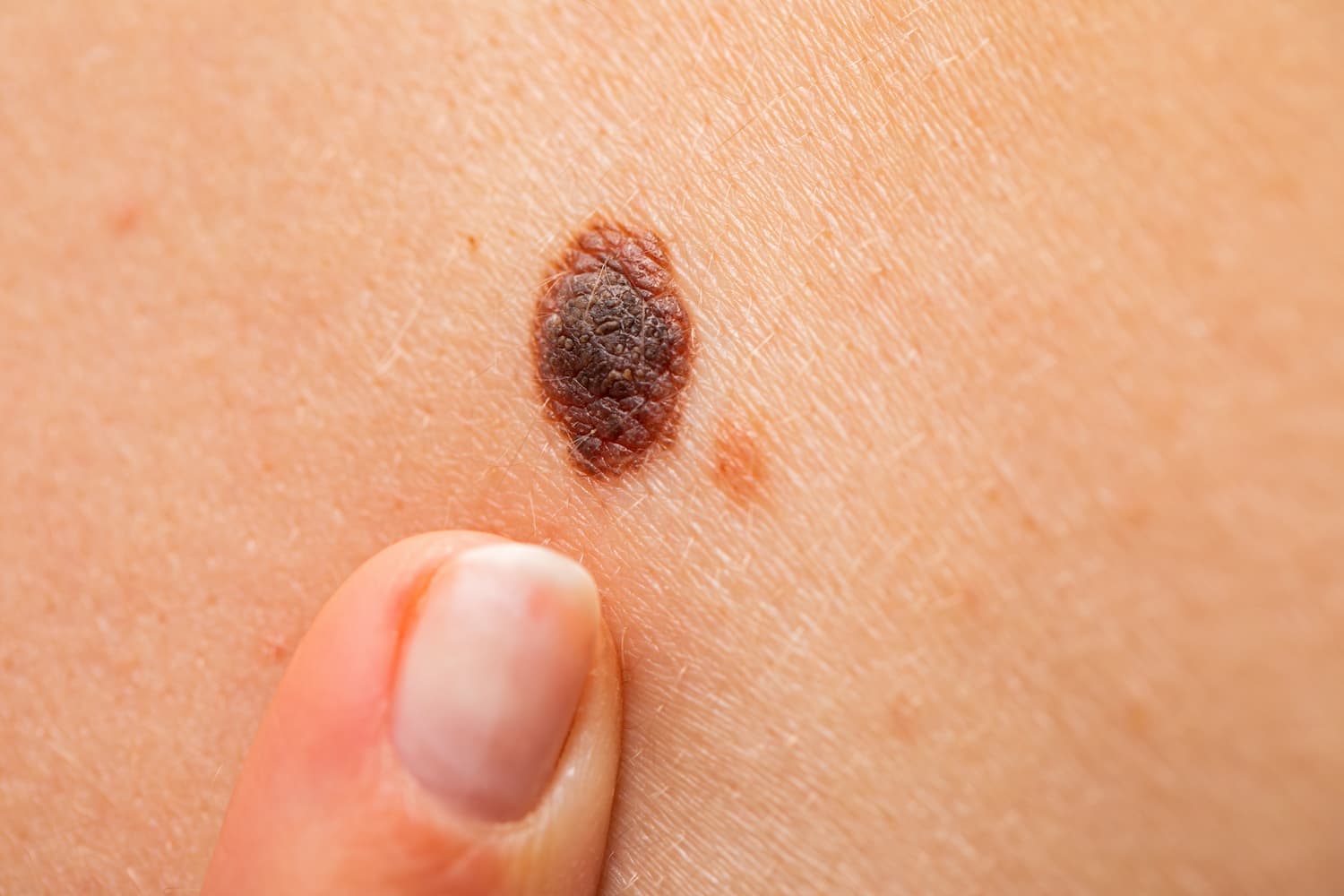
## 5. Clinical Features
- Size: usually <6 mm (bigger lesions = higher suspicion).
- Symmetry, regular borders, uniform color → benign.
- Common on face, trunk, extremities.
- May involute with age (especially intradermal nevi).
---
## 6. Risk of Malignancy
- **Acquired small nevi**: very low risk.
- **Dysplastic (atypical) nevi (이형성 모반)**: architectural disorder, cytologic atypia → moderate melanoma risk.
- **Congenital giant nevi (>20 cm)**: lifetime melanoma risk up to 5–10%.
---
## 7. Management
- **Observation**: Most nevi need no treatment.
- **Excisional biopsy**: Gold standard if suspicious features present (ABCDE rule).
- **Laser treatment**:
- Cosmetic only, not diagnostic.
- Must rule out atypia/melanoma first (biopsy if uncertain).
- **Nd:YAG (532/1064 nm)** for junctional/epidermal pigment.
- **CO₂ laser** for raised dermal nevi.
---
## 8. Clinical Pearls
1. **Never laser a nevus without diagnostic certainty.** Histology is lost after ablation.
2. **Maturation with depth** = hallmark of benignity. Lack of maturation = melanoma.
3. **“Ugly duckling sign”**: One nevus that looks different from the others warrants biopsy.
4. **Patient counseling**: Educate about self-monitoring, especially for dysplastic nevi or family history of melanoma.