### Date : 2025-02-17 23:41
### Topic : 1.3 Hair Follicle Anatomy
----
# **1.3 Hair Follicle Anatomy**
The **hair follicle** is a **complex mini-organ** embedded in the skin, responsible for **hair production, growth cycle regulation, and regeneration**. Understanding its detailed structure is crucial for **hair transplantation, diagnosing hair disorders, and optimizing graft survival**.
---
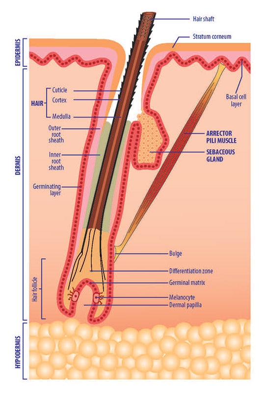
## **1️⃣ Overview of Hair Follicle Structure**
The hair follicle consists of three main parts:
1. **Hair Bulb (Deepest part – Growth center)**
2. **Hair Root (Middle section – Anchoring zone)**
3. **Hair Shaft (Visible part above skin)**
### **Layers of the Hair Follicle**
The follicle is composed of **concentric layers** that support hair growth:
|**Layer**|**Function**|
|---|---|
|**Inner Root Sheath (IRS)**|Guides hair growth, dissolves at surface|
|**Outer Root Sheath (ORS)**|Contains stem cells, regenerates follicle|
|**Connective Tissue Sheath (CTS)**|Provides structural support|
---
## **2️⃣ Hair Follicle Zones & Clinical Importance**
### **A. Hair Bulb (The Root of Growth)**
- Located at the **deepest part** of the follicle (~3.5–5.0 mm in the scalp).
- Contains **dermal papilla (growth regulator)** and **matrix cells**.
💡 **Clinical Relevance:**
✔ **Dermal Papilla (DP)** → The **"brain" of the follicle**, responsible for **hair growth & cycling**.
✔ Must be **extracted intact during FUE/FUT**—**damage to DP = no regrowth**.
✔ **Melanocytes in the bulb** produce hair pigment—dysfunction causes **gray hair**.
---
### **B. Outer Root Sheath (ORS) – The Regeneration Center**
- Houses **stem cells** in the **bulge region** (near the arrector pili muscle).
- Provides **protection & mechanical strength** to the follicle.
💡 **Clinical Relevance:**
✔ **Essential for graft survival**—preserving ORS ensures **follicle regrowth after transplant**.
✔ **DHT-sensitive in androgenetic alopecia (AGA)** → Leads to follicle miniaturization.
✔ **Rich in keratinocytes**—important for wound healing after extraction.
---
### **C. Inner Root Sheath (IRS) – Hair Molding Layer**
- Forms a **temporary scaffold** around the hair shaft.
- Degenerates before the hair emerges from the skin.
💡 **Clinical Relevance:**
✔ Helps **align and shape the hair shaft**—disruptions can lead to **curved or brittle hair**.
✔ In curly hair (Afro-textured), the IRS is **more pronounced**—affects FUE extraction angles.
---
### **D. Dermal Papilla (DP) – The Growth Command Center**
- Located at the **base of the hair bulb**.
- Contains **fibroblasts, capillaries, and growth factors**.
- Regulates **hair cycle transitions (anagen, catagen, telogen).**
💡 **Clinical Relevance:**
✔ **Must be preserved in transplants**—a **DP-less graft will not grow**.
✔ **Target for stem cell therapy & hair cloning research**.
✔ Plays a **role in hair thickness & speed of regrowth**.
---
### **E. Sebaceous Gland – The Follicle’s Lubricant**
- Produces **sebum (oil)** to keep hair soft.
- Located in the **mid-dermis**, connected to the hair follicle.
💡 **Clinical Relevance:**
✔ Helps prevent **graft desiccation (drying out) after extraction**.
✔ Overactive sebaceous glands contribute to **seborrheic dermatitis, folliculitis, acne**.
---
### **F. Arrector Pili Muscle – The "Goosebump" Muscle**
- Small muscle attached to the hair follicle.
- Contracts in response to **cold, fear, or stress** → Causes "goosebumps."
💡 **Clinical Relevance:**
✔ Not critical in transplants but plays a **role in scalp microcirculation**.
✔ Loss of attachment can **slightly affect follicle positioning after FUE**.
---
## **3️⃣ Hair Shaft (Emerging Hair)**
Once the follicle produces hair, the **visible part** is called the **hair shaft**, composed of **keratin**.
### **Three Layers of the Hair Shaft**
4. **Medulla (Innermost)** – Soft keratin, present in thick hair.
5. **Cortex (Middle Layer)** – Determines **strength, thickness, color**.
6. **Cuticle (Outermost Layer)** – Overlapping cells that **protect hair**.
💡 **Clinical Relevance:**
✔ **Weak cuticle = split ends, hair breakage**.
✔ **Hair straighteners & dyes damage the cortex**, leading to brittle hair.
---
## **4️⃣ Hair Follicle Classification**
Hair follicles are classified based on their **grouping pattern and density**.
|**Follicular Unit Type**|**Hair Count Per Follicle**|**Location**|
|---|---|---|
|**Single Hair Follicle**|1 hair|Hairline, eyebrows|
|**Follicular Unit (FU)**|2–3 hairs|Most of the scalp|
|**Multi-Follicular Unit (MFU)**|4–6 hairs|Occipital donor zone|
💡 **Clinical Relevance:**
✔ **Hairline transplants use single FUs for a natural look**.
✔ **Crown and vertex areas require 2–3 hairs per FU for density**.
---
## **5️⃣ Hair Follicle Depth Variations by Scalp Region**
|**Scalp Area**|**Average Follicle Depth**|
|---|---|
|**Frontal Hairline**|3.5 mm|
|**Temporal Area**|3.0 mm|
|**Mid-Scalp**|4.0 mm|
|**Crown (Vertex)**|4.5 mm|
|**Occipital Donor Zone**|4.5–5.0 mm|
💡 **Clinical Relevance:**
✔ **FUE punch depth must be adjusted based on scalp region**.
✔ Over-punching beyond the **bulb depth (~4.5mm)** can lead to **excessive bleeding, scarring**.
---
## **📌 Key Takeaways**
✅ **The hair follicle is a complex structure, NOT just a simple root.**
✅ **Dermal papilla must be preserved for transplant success.**
✅ **Outer root sheath contains stem cells for follicle regeneration.**
✅ **Hair follicle depth varies across the scalp (3.0 – 5.0 mm).**
✅ **FUE punch depth should not exceed the hypodermis (>4.5 mm) to avoid damage.**
---